
Figure 1.
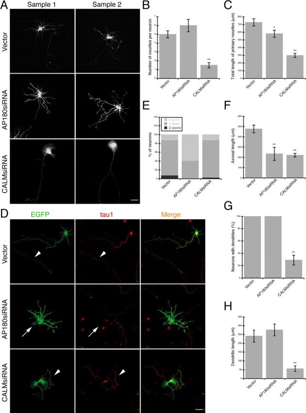
A–H, Reduction of AP180 and CALM leads to different growth defects in hippocampal neurons. A, Confocal images of representative embryonic hippocampal neurons expressing AP180siRNA or CALMsiRNA. Scale bar, 10 μm. Additional examples are shown in supplemental Figure S4A, available at www.jneurosci.org as supplemental material. B, The number of neurites per neuron is not significantly changed in neurons expressing AP180siRNA but is reduced in neurons expressing CALMsiRNA. n = 60 neurons in each group; **p < 0.001. C, Total neurite length is reduced in neurons expressing AP180siRNA and CALMsiRNA. n = 60 neurons in each group; *p < 0.05, **p < 0.001. D, Immunolabeling of the axonal marker tau1 (red) in neurons coexpressing EGFP (green) and the indicated siRNA. Arrowheads indicate the tau1-labeled axon. Arrows mark an AP180siRNA neuron in which the axon is absent. Scale bar, 10 μm. E, Proportion of neurons containing tau1-labeled axons as shown in D. n = 60 neurons in each group. F, The mean length of axons is reduced by both AP180siRNA and CALMsiRNA. Vector, n = 45 neurons; AP180siRNA, n = 20 neurons; CALMsiRNA n = 45 neurons; **p < 0.001. G, Quantification of neurons containing MAP2-labeled dendrites. n = 45 neurons in each group; **p < 0.001. H, The total length of dendrites is reduced in the CALMsiRNA-expressing neurons. Vector and AP180siRNA, n = 45; CALMsiRNA, n = 12; **p < 0.001. Data represent means ± SEM in B, C, F–H.