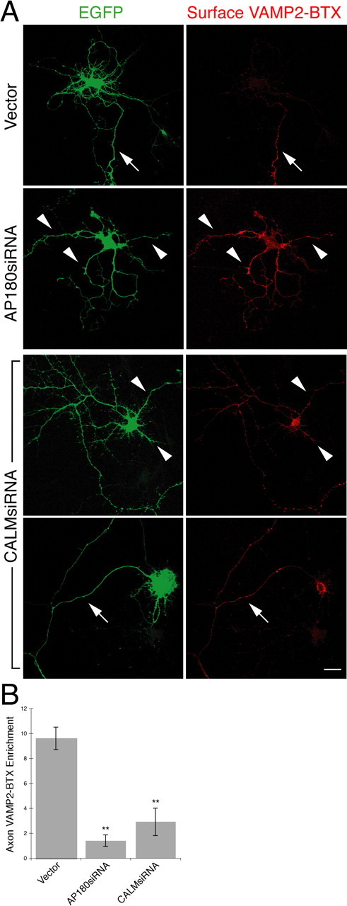
Figure 9.

Knockdown of AP180 and CALM disturbs VAMP2 distribution. Live neurons coexpressing EGFP (green), VAMP2-BTX, and the siRNA were labeled with Alexa555-BTX (red) at 4°C. The neurons were then fixed and confocal images were acquired. A, Surface VAMP2-BTX in the AP180siRNA neurons or the CALMsiRNA neurons. In the control neuron, surface VAMP2-BTX is detected only on the axon (arrow). In the AP180siRNA neuron, surface VAMP2 becomes detectable on all neurites (arrowheads). Among the CALMsiRNA neurons, some of them have a seemingly unaltered morphology (top), but they express surface VAMP2 on all neurites (arrowheads); others have most of their dendrites missing (bottom), and surface VAMP2 is brightly labeled on the axon. Scale bar, 10 μm. B, Quantification of VAMP2 axon enrichment. Data represent average intensity of BTX fluorescence in the longest neurite divided by that in all other neurites. **p < 0.001, n = 30 neurons for the control and AP180siRNA; n = 15 neurons for CALMsiRNA.
