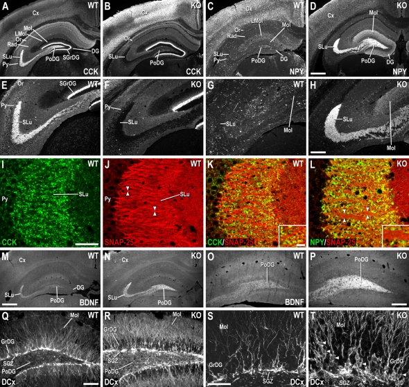
Figure 6. SNAP-25, CCK, NPY, BDNF, and DCx Immunoreactivity in the Hippocampal Formation of Adult Neo-excised SNAP-25b Deficient and WT Mice.
(A) CCK-ir in HF of WT mice, shown at higher magnification in (E). In SNAP-25b deficient mice (B), CCK-ir disappears in PoDG and SLu, higher magnification in (F). (C) NPY-ir in WT HF, shown at higher magnification in (G). (D) Increased NPY-ir in PoDG, Mol, and SLu of SNAP-25b deficient (KO) mice, the latter at higher magnification in (H). Confocal micrographs of double-staining experiments taken from WT (I-K), and neo-excised SNAP-25b deficient (L) mice. CCK- (I) and SNAP-25-ir (J) in fibers/terminals of SLu in WT mice. Arrowheads in (J) point to strong SNAP-25+ fibers. (K) Double-staining of CCK- (green) and SNAP-25-ir (red) in WT SLu. (L) NPY-ir, lacking in fibers/terminals of WT SLu, is highly expressed in neo-excised SNAP-25b deficient mutants. In homozygous neo-excised SNAP-25b deficient mice, SNAP-25-ir fibers in SLu are often thicker (arrowheads in (L) compared to WT mice [(L) cf. (J,K)]. Insets in (K,L) show respective photomicrographs at higher magnification. (M) BDNF-ir in WT HF, the PoDG, and SLu shown at higher magnification in (O). (N) Increased BDNF-ir in PoDG and SLu of mutant mice, the DG shown at higher magnification in (P). (Q) Strong DCx+ cell bodies in SGZ of WT DG, shown at higher magnification in (S). (R) Increased number of cell bodies randomly distributed in SGZ and granular cell layer of neo-excised SNAP-25b deficient mutants, with higher levels of DCx+ fibers in Mol, shown at higher magnification in (T). Arrowheads in (T) point to DCx+ cell bodies in the granular cell layer. Cx, cortex; DG, dentate gyrus; LMol, lacunosum Mol; Mol, molecular layer of DG; Or, oriens layer of HF; PoDG, polymorph layer of DG; Py, pyramidal layer of HF; Rad, stratum radiatum of HF; SGrDG, supragranular layer of DG; SGZ, subgranular zone of DG; SLu, stratum lucidum of HF. Scale bars: (D = A–C = 500 µm), (H = E–G = 200 µm), (I = J–L = 50 µm), (K, inset = L inset = 10 µm), (M = N = 500 µm), (O = P = 200 µm), (Q = R = 100 µm), (S = T = 50 µm). WT (n = 2), SNAP-25b mutant mice (n = 2).

