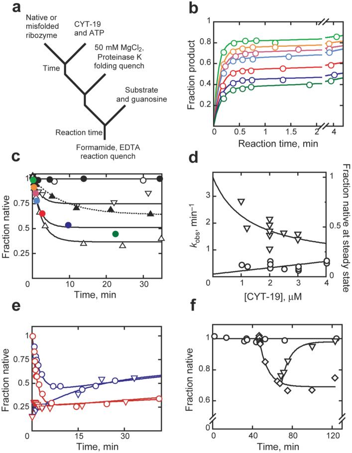
Figure 1.
Unfolding of native and misfolded Tetrahymena ribozyme. a, Reaction scheme. b, Substrate cleavage after incubation with 1 mM Mg2+, 2 μM CYT-19 and 2 mM ATP-Mg2+ for 0.25 (orange), 0.67 (red), 1 (cyan), 2.5 (magenta), 9.5 (blue), or 22 min (dark green). (Light green), no CYT-19. c, Native ribozyme unfolding (1 mM Mg2+). CYT-19 was 1 μM (∇), 2 μM (solid colored circles), or 3 μM without (σ) or with 2 mM ATP-Mg2+ (∆). Colored circles show burst amplitudes from corresponding curves (panel b). ○, no CYT-19; ●, 2 μM CYT-19, 2 mM ATP-Mg2+, 5 mM Mg2+. d, Rate constant (○) and steady-state value (∇) vs CYT-19 concentration. e, Approach to steady state from native (○) or misfolded (∇) ribozyme with 1.2 μM (blue) or 2 μM (red) CYT-19. f, Refolding to the native state (∇) after unfolding by CYT-19 (◇) and inactivation by proteinase K. ○, no CYT-19.

