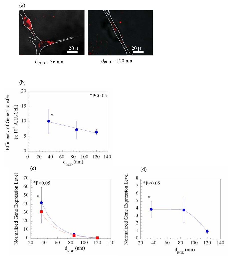
Figure 3.
Gene delivery regulated with the distance between islands of RGD peptides (dRGD), as the total density of RGD peptides was kept constant at 6 × 10⁹/mm2. Microscopic images show the decrease in the cellular uptake of the rhodamine-labeled pDNA condensates with dRGD (a). White lines in photomicrographs represent borders of cells adhered to the gel matrix. The efficiency of gene transfer quantified from photomicrographs was linearly decreased with dRGD (R2 = 0.96) (b). The gene expression level (● in c) and the gene expression level normalized to the average surface area of cells (■ in c) exponentially decreased with dRGD (R2 = 1). Increasing dRGD also down-regulated the gene expression level in cells cultured within the 3D gel matrix (d). Differences in the values in (b) for cells cultured on gels presenting the largest dRGD (~ 120 nm) versus those on gels presenting the smallest dRGD (~ 36 nm) were statistically significant (p < 0.05). The gene expression levels in (c) and (d) were normalized to the value attained from cells cultured on gels presenting dRGD of 120 nm. Data points and error bars represent the mean and standard deviation from four independent experiments.

