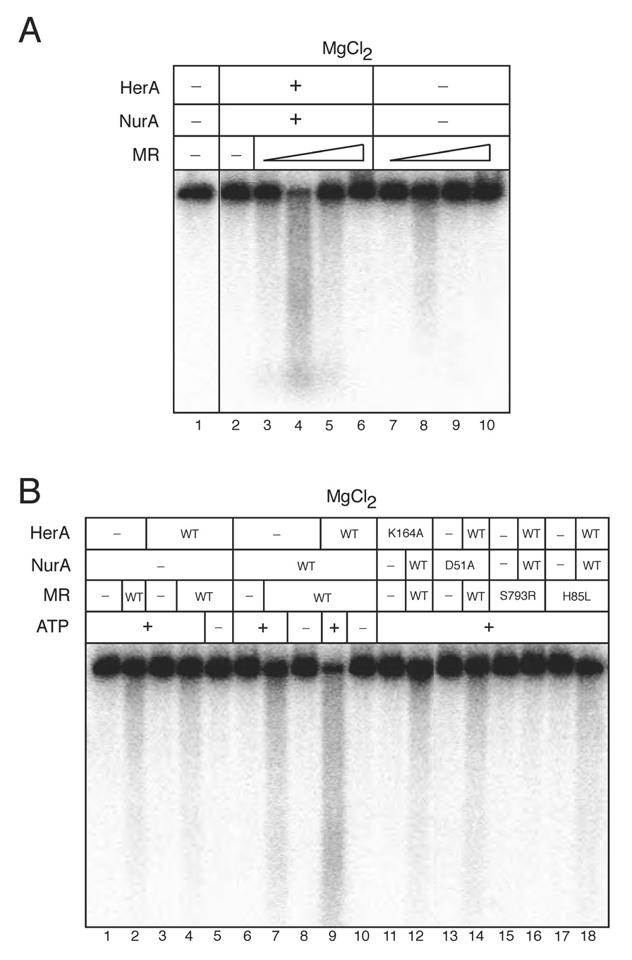
Figure 1. HerA and NurA degrade dsDNA cooperatively with Mre11/Rad50 in magnesium.
(A) Nuclease assays were performed using 2.5 kb internally [32P]-labeled dsDNA. Reactions contained 2.7 nM wild-type HerA and 19.2 nM wild-type NurA, 5 mM MgCl2, 1 mM ATP, and 100 mM NaCl. Wild-type MR complex was included in the reaction at 0.3, 3.3, 33, and 330 nM. HerA molar concentrations are given as a hexamer of 370 kDa, NurA concentrations are given as a monomer of 52 kDa, and MR complex is given as a 2:2 stoichiometric complex of 306 kDa.
(B) Reactions performed as in (A) with HerA wild-type or K164A, NurA wild-type or D51A, and 3.3 nM MR wild-type, M(H85L)R, or MR(S793R) as indicated in 80 mM NaCl.

