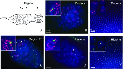
Figure 2.—
FISH pairing assay in germaria and embryos of control w1118 flies. (A) Cartoon diagram of the germarium. Pairing was assayed collectively in C(3)G staining nuclei of regions 2a and 2b. (B) Paired dodeca foci (green), partially separated between a strand of SC, indicated by immunofluorescence (red) to C(3)G in region 2a. (C) A single focus (green) using a region 25 BAC probe indicating pairing in a meiotic nucleus of 2a. (D) A single focus (green) using a histone cluster probe indicating pairing in a meiotic nucleus of 2a. (E and F) FISH in early embryos. FISH experiments were performed on 2- to 4-hr-old embryos. Pairing was assayed in embryos that had completed cellularization and just begun gastrulation (∼3 hr). (E) Dodeca probe. (F) Histone cluster probe. In all panels blue indicates DAPI. Each panel is a single z-section.

