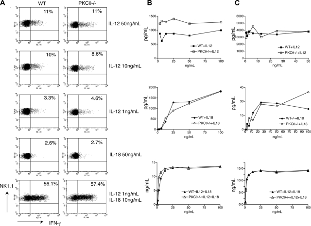
Figure 4.
Cytokine-induced IFN-γ production is PKC-θ–independent. (A) PKC-θ−/− and WT splenocytes were stimulated ex vivo with the indicated doses of IL-12 and/or IL-18. Intracellular IFN-γ content was measured by FACS, gating on NK1.1+CD3−CD19− NK cells. (B) IL-2–cultured PKC-θ−/− and WT NK cells were stimulated with the indicated doses of IL-12, IL-18, or IL-12 and IL-18 for 24 hours. IFN-γ released in cell culture supernatants was determined by CBA or ELISA. IFN-γ concentrations are indicated in picograms per milliliter (IL-12, IL-18) or nanograms per milliliter (IL-12 + lL-18). (C) Splenocytes from PKC-θ−/− and WT mice were cultured for 1 week in IL-15 and then stimulated for 24 hours with the indicated doses of IL-12, IL-18, or IL-12 and IL-18. IFN-γ released in cell culture supernatants was determined by CBA or ELISA. IFN-γ concentrations are indicated in picograms per milliliter (IL-12, IL-18) or nanograms per milliliter (IL-12 + lL-18).

