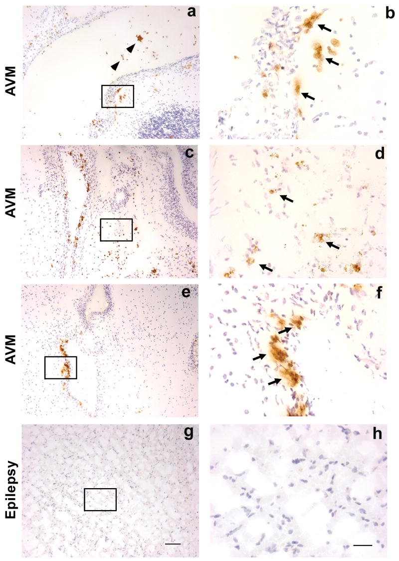
Figure 1.
Immunohistochemical staining of neutrophil marker MPO on sections from un-ruptured and non-embolized brain AVM tissue (figure a–f) and epilepsy (figure g–h) control tissue. Brown color, indicated by arrows, is positive MPO signal, and blue color is the counter staining with hematoxylin of nuclei. Figure a showed that neutrophils were present in the vascular wall and lumen of AVMs. Figure b was photographed under higher power from selected area of the figure a, indicted by rectangle. Figure c showed neutrophils were present in the vascular wall as well as parenchymal tissue of AVMs, and figure d was photographed under higher power from selected area of c, indicted by rectangle. Figure e showed that neutrophils were predominantly present in the vascular wall, and image f showed selected area from e under higher magnification. Neutrophils were absent in cortical tissue of epilepsy patient (figure g and h). Representative images from 3 AVM patients and an epilepsy patient; size bar for a, c, e, and g: 100μm; size bar for b, d, f, and h: 25μm

