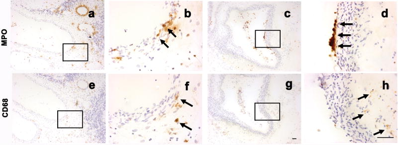
Figure 3.
Co-localization of MPO and CD68 signal. Adjacent AVM tissue sections were stained with MPO, or CD68 marker. Brown color, shown by arrows, indicates positive MPO, or CD68 immunoreactivity. Cell nuclei appear blue with hematoxylin counter staining. Figure a and e showed that neutrophils and macrophages were present in proximity at the same vessels. Figure b and f were photographed under higher power from selected area, indicted by rectangle, of a, e respectively. Figure c and g showed that neutrophils and macrophages were present at different locations: neutrophils were mainly present at the inner wall; macrophages were mainly located at the outer vascular wall. Figure d and h were photographed under higher power from selected area, indicted by rectangle, of c, g respectively. Size bar for a, c, e, and g: 50μm; size bar for b, d, f, and h: 50μm

