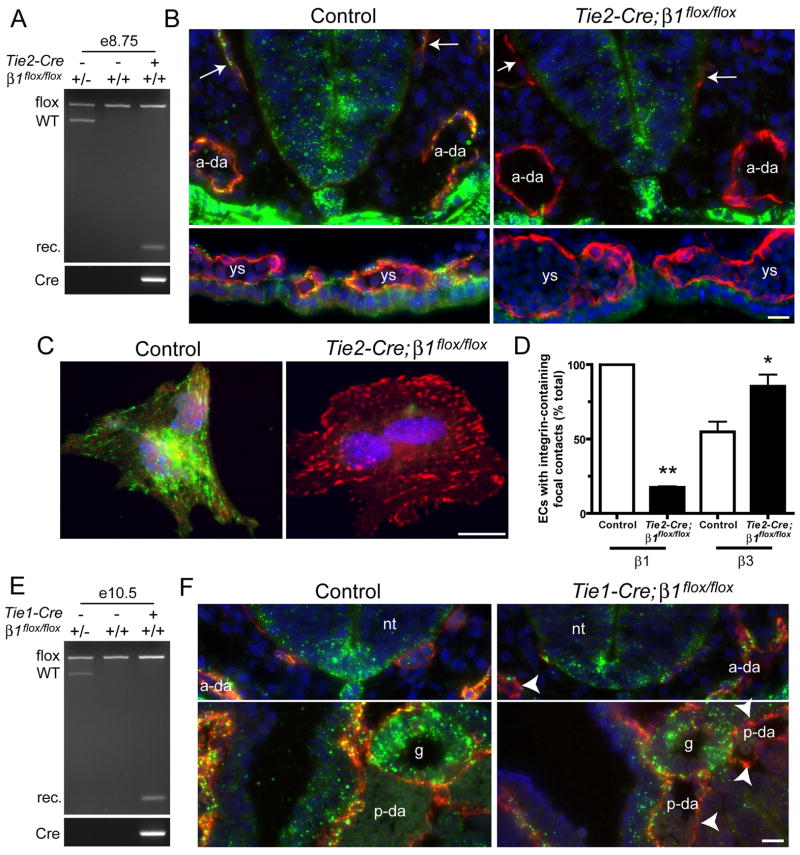
Figure 1. Tie2- and Tie1-Cre mediate efficient deletion of β1 integrins in embryos.
Gene deletion analyses of Tie2- (A–D) and Tie1- (E,F) mutants. (A) Genomic PCR analysis of e8.5 embryos demonstrates recombination (rec.) of β1 in embryos carrying Tie2-Cre and two floxed alleles of β1. (B) e8.5 cryosections were stained with anti-CD31 (red), anti-β1 integrin (Ha2/5, green), and DAPI (blue). (C) Collagenase-dissociated e8.5 embryonic cells were plated onto FN and stained with anti-β1 (HMβ1-1, green), anti-β3 (red), and DAPI (blue). EC identity in C was determined by co-staining with anti-CD31 (not shown). (D) Focal contacts in isolated ECs. Bars are means + SEM of 2 (β1) or 3 (β3) experiments. **p < 0.01 by one sample T-Test and *p < 0.05 by Student’s T-test. (E) Genomic PCR analysis of e10.5 embryos from Tie1-Cre matings. (F) e9.5 cryosections were stained with anti-CD31 (red), anti-β1 integrin (green), and DAPI (blue). Arrows, primary head veins; arrowheads, β1-negative endothelium; ys, yolk sac blood islands; a-da, anterior dorsal aortae; p-da, posterior dorsal aortae; nt, neural tube; g, gut; ve, visceral endoderm. Also see Fig. 3F for diagram of embryonic structures. Bars, 20 μm.

