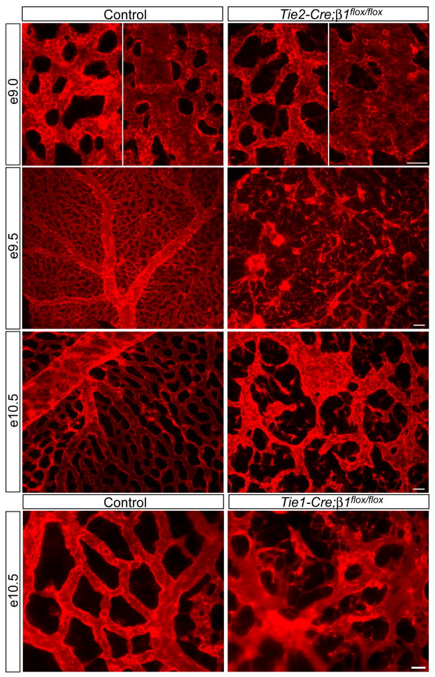
Figure 2. Endothelial deletion of β1 integrins causes EC disorganization leading to cardiovascular defects and lethality at midgestation.
Whole mount anti-CD31 immunostained yolk sacs at the indicated embryonic stages. The left panels of e9.0 are capillary regions and the right panels are regions apparently undergoing arteriovenous remodeling. Note the disconnected capillaries and overall vascular disorganization in all mutants. Images are representative of at least 5 control/mutant embryo pairs at each stage. Bars, 100 μm (e9.5), 50 μm (e9.0 and e10.5, Tie2-Cre), and 25 μm (Tie1-Cre).

