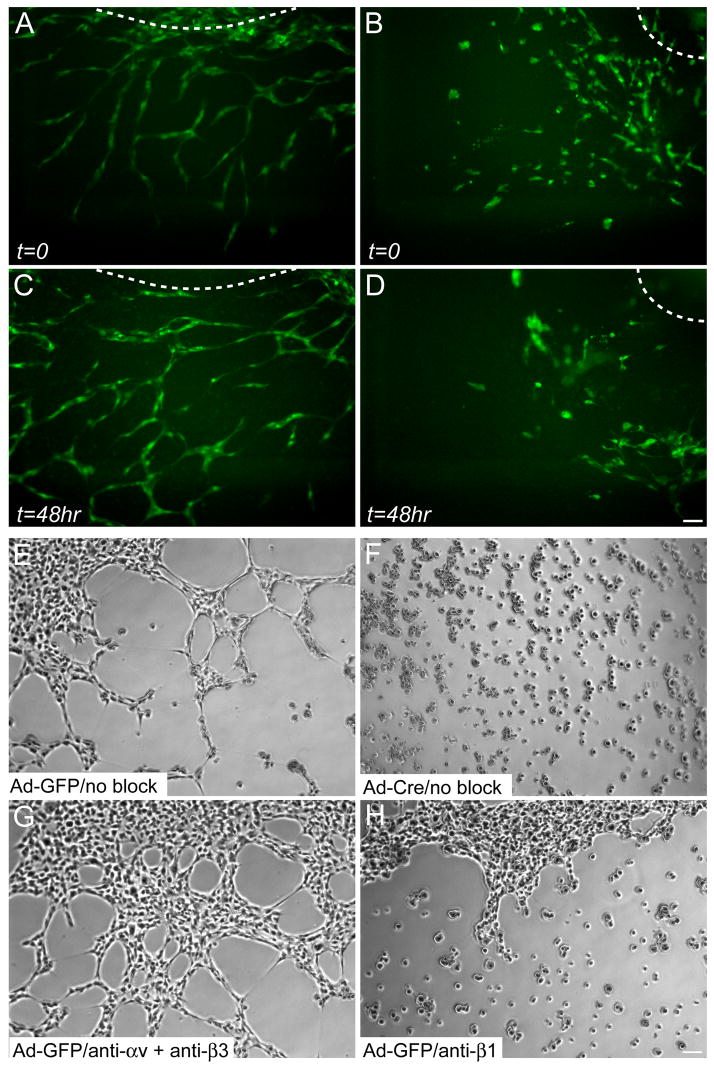
Figure 5. EC-autonomous role for β1 integrins in angiogenic remodeling.
Still images of control (A,C) and Tie2-Cre mutant (B,D) P-Sp explant cultures at the indicated timepoints. GFP-positive ECs were visualized by a Tie1-GFP transgene. Aberrant EC clusters are evident in Tie2-Cre mutant explants, but not in control explants. The OP9 feeder cells, not visible in the fluorescence images, were a confluent monolayer on top of which the ECs grew out from the P-Sp. The dashed lines indicate the approximate location of the P-Sp explants. See also supplemental online movies. (E–H) Capillary morphogenesis of embryonic β1flox/flox ECs immortalized with PyMT and subsequently infected with adenovirus (Ad), in the absence of any antibodies (E,F) or in the presence of anti-αv plus anti-β3 (G) or anti-β1 (H) function-blocking integrin antibodies. Phase contrast images were captured after 4 hr in culture. Bars, 100 μm.

