
Figure 11.
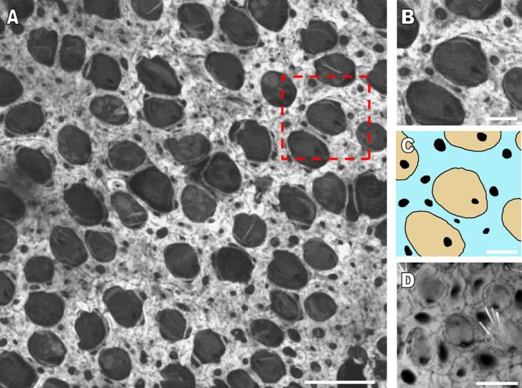
The width of circumferential F-actin bands increases in the utricular supporting cells of developing humans and other murine balance organs. A) A high resolution confocal image of the sensory epithelium of a utricle stained with phalloidin from a 60 year-old human cadaver shows the extent to which the F-actin bands cover supporting cell apical areas. Scale bar, 15 μm. B and C) A zoomed in region of the sensory epithelium (box outlined with red dashes in A) shows the location of the bands in supporting cells (blue) in relation to the cuticular plates of hair cells (tan). Very little area remains free of F-actin in the supporting cell apical areas. Scale bar in B and C, 5 μm. D) A high resolution confocal image of the sensory epithelium in the cristae ampullaris from a postnatal day 120 mouse shows that the circumferential F-actin bands of supporting cells have almost an identical morphology to that seen in the utricles of adult mice. Scale bar, 5 μm.