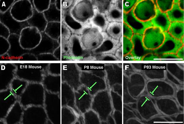
Figure 2.
A space between the circumferential F-actin bands of adjacent supporting cells which becomes resolvable as mice age is at the level of the adherens junctions. A-C) Images of N-cadherin immunohistochemistry (red), phalloidin staining (green), and their overlaid image show there is a visible gap between the F-actin bands at the intercellular adherens junction joining supporting cells in a postnatal day 8 mouse utricle. These frames are a single 0.3 μm thick confocal slice at the z-depth where the fluorescence intensity of N-cadherin immunohistochemistry was at its highest. A large accumulation of F-actin was visible at this depth. Scale bar for A-C in C, 5 μm. D-F) High magnification images of cells from the nonsensory epithelium from Fig. 1A show the width of circumferential F-actin bands increases in cells of the nonsensory epithelium as mice age, but not to the extent that it does in supporting cells of the sensory epithelium. Scale bar for D-F in F, 10 μm. (A magenta-green version of this figure is provided in the supplementary material)

