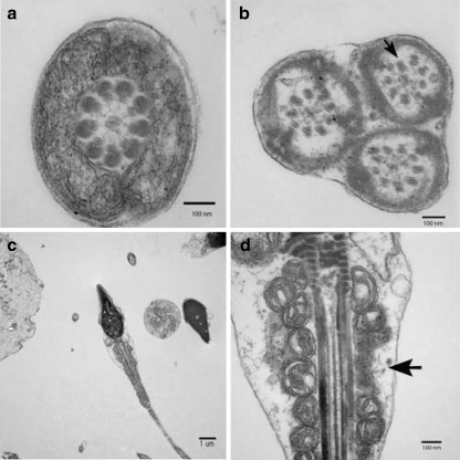
Fig. 2.
Transmission electron micrograph of various spermatozoa from the patient with PCD. A) A cross section through a mid-piece region of flagellum. B) Three axonemes surrounded by the same outer membrane. Notice the partial axonemal disruption (Arrow). C) Transverse section of a spermatozoon. D) Same spermatozoon flagellum at a higher magnification. Notice the disruption of the mitochondrial sheath (Arrow)

