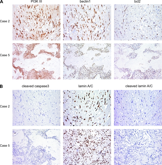
Figure 4.
Immunohistochemical analysis of two imatinib-treated GISTs, showing residual cellularity b (case 2) and c (case 5). (A) Expression of autophagy-related proteins. Both cases show cytoplasm decoration for PI3KIII, beclin1, and bcl2. (B) Expression of apoptosisrelated proteins. Both cases show nuclear immunoreactivity for full-length lamin A/C and no immunostaining for cleaved/activated caspase 3 or cleaved lamin A/C.

