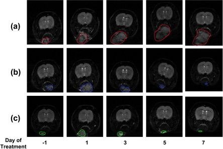
Figure 2.
Submental tumor growth. Sequential T2-weighted MRI images of a slice through a control (A), a 5FC-treated (B), and a 5FC and radiation-treated (C) yCD-expressing SCCVII tumor. Images were acquired 1 day before therapy and 1, 3, 5, and 7 days after therapy. The submental tumors are indicated by the red, blue, and green regions of interest, respectively.

