
Figure 2. M3 effective coverage maps for deficits in recognition and naming, thresholded at p<0.001.
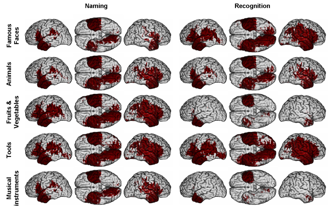
Red color indicates where a significant difference between the number of subjects with a lesion and a deficit and the number of subjects with a lesion and no deficit can potentially be detected. Each row of brains corresponds to a given category of concrete entities. The three first columns correspond to results related to naming performance and the three last columns to results related to recognition performance. In each group of three columns, three views of the brain are presented: left lateral (left), ventral view of both hemispheres (middle), and right lateral (right). (See Supplementary Material online for parallel PM3 and reduced model maps).