
Figure 3.
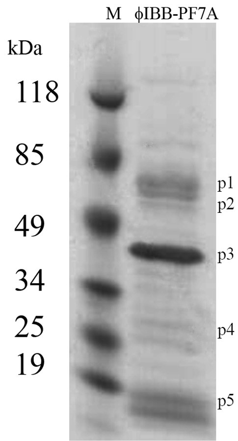
SDS-polyacrylamide gel electrophoresis analysis of phage ϕIBB-PF7A structural proteins. Phage lysate was mixed with Laemmli buffer containing SDS, boiled for 10 min, and loaded on a 4–20% gradient gel that was electrophoresed with Tris-glycine running buffer. Lane M: molecular weight marker. p1 to p5 mark sizes of typical T7 phage structural proteins. Further explanations in the Results section.