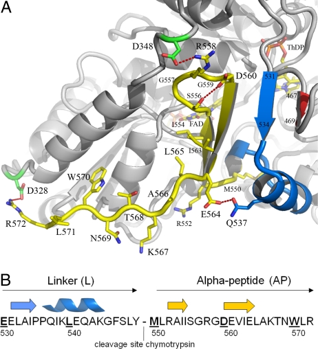
Fig. 3.
Structure of the C-terminal membrane-binding domain of EcPOX. (A) Diagram representation of EcPOX in gray with the C terminus highlighted: blue, linker region (residues 531–549); yellow, alpha-peptide part (residues 550–572). The cofactors ThDP and FAD, and selected amino acid side chains are shown in stick representation. Selected hydrogen-bonding and electrostatic interactions are indicated. (B) Primary sequence and secondary structure assignment of the C-terminal domain.

