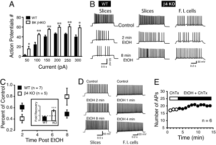
Fig. 3.
EtOH-mediated decrease of MSN excitability exhibits tolerance in KO, but not WT mice. (A) Number of APs recorded from WT (filled columns) and KO (open columns) MSNs following a series of incremental (50 pA) current steps (50–300 pA). (B) Representative action potential trains evoked in a slice preparation (Slices) by a single 100 pA current step in WT (Left) and KO (Middle) mice before (control) and after 50 mM EtOH exposure (2 or 8 min). Two minutes after EtOH, the number of APs is smaller in both WT and KO mice. While KO mice MSN excitability partially recovers 8 min after EtOH exposure (Bottom Trace; Right), WT neuronal excitability remains depressed (Left, Bottom Trace). Results obtained in slices were reproduced on freshly isolated neurons from KO mice (Right; β4 KO/F.I. cells). (C) Averaged change in action potential number recorded in MSNs in slices and freshly isolated after 2 or 8 min EtOH exposure, presented as percent of control before EtOH exposure in MSNs from WT and β4 KO striatal slices; 5/7 neurons were ethanol sensitive and developed tolerance in β4 KO MSNs, whereas 7/9 MSNs from WT were ethanol sensitive and did not develop tolerance (*P < 0.05). The Inset shows the ratio of AP number at 2 and 8 min reported as fold recovery from ethanol; value below the broken bar indicates a further decrease of APs number at 8 min compared to 2 min EtOH (solid column; WT), while value above the line indicate a recovery (KO); F1,10 = 27.6, P < 0.001. (D) 100 nM ChTx blocks EtOH effects on striatal MSNs AP patterns in slices (Left) and freshly isolated neurons (F.I cells, Right). (E) Number of APs measured every minute before (open circles) and during 50 mM EtOH exposure (solid circles) in presence of 100 nM ChTx. Data from slices and freshly isolated MSNs were combined.

