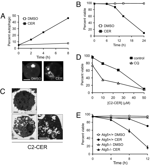
Fig. 1.
Ceramide triggers homeostatic autophagy. (A) Cells expressing GFP-LC3 were treated with DMSO or 50 μM C2-cer for the indicated intervals. Representative cells at 4 h are shown. Scale bar, 10 μm. (B) Viability of cells treated with DMSO or with 50 μM C2-cer. (C) Cells expressing Bcl-XL treated with DMSO or 50 μM C2-cer for 24 h were examined by electron microscopy. Scale bars, 2 μm or 1 μm. (D) Viability of cells treated for 24 h with C2-cer with or without 10 μM CQ. 3-MA was toxic even in the absence of ceramide (data not shown). (E) Viability of wild-type and Atg5−/− MEFs treated with 20 μM C2-cer in 1% FCS. Error bars, SD.

