. 2008 Aug 28;61(11):1160–1167. doi: 10.1136/jcp.2008.055335

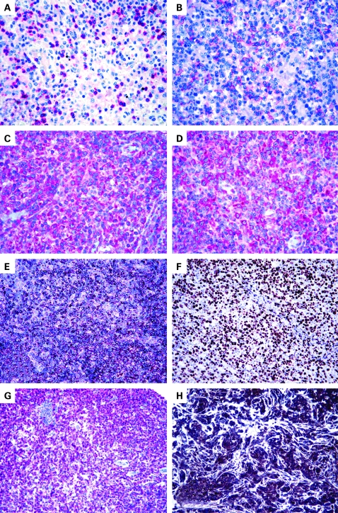
Figure 1. (A) Lymphomatous cells do not express CD4; however, CD4 is detected in some reactive small lymphocytes (alkaline phosphatase anti-alkaline phosphatase (APAAP) technique, Gill’s haematoxylin nuclear counterstaining, ×250). (B) Partial CD30 expression; it should be noted that the tumour has no anaplastic morphology (APAAP technique, Gill’s haematoxylin nuclear counterstaining, ×250). (C) Positivity for platelet-derived growth factor receptor α (PDGFRα) (APAAP technique, Gill’s haematoxylin nuclear counterstaining, ×400). (D) PDGFRα is phosphorylated (APAAP technique, Gill’s haematoxylin nuclear counterstaining, ×400). (E) CXCL13 expression by neoplastic elements in angioimmunoblastic T cell (EnVision+ technique, Gill’s haematoxylin nuclear counterstaining, ×100). (F) Ki-67 marking exceeds the 80% value (EnVision+ technique, Gill’s haematoxylin nuclear counterstaining, ×200). (G) CD52 positivity in a peripheral T cell lymphoma, not otherwise specified (APAAP technique, Gill’s haematoxylin nuclear counterstaining, ×100). (H) Strong expression of vascular endothelial growth factor in an angioimmunoblastic T cell lymphoma (EnVision+ technique, Gill’s haematoxylin nuclear counterstaining, ×200).

Figure 1