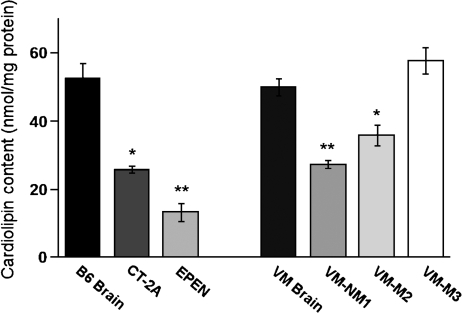
Fig. 2.
CL content in mitochondria isolated from mouse brain and brain tumors. Mitochondria were isolated as described in Materials and Methods. Values are represented as the mean ± SD of three independent mitochondrial preparations from brain or tumor tissue. Asterisks indicate that the tumor values differ significantly from the B6 or the VM brain values at the * P < 0.01 or ** P < 0.001 levels as determined by the two-tailed t-test.

