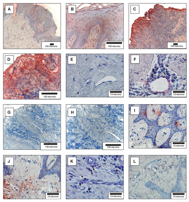
Figure 1.

SDF-1α and CXCR4 expression in normal and periodontal diseased human gingiva. Human gingival specimens were stained for the expression of SDF-1α immunohistochemistry (red) and counterstained by hematoxylin (blue). Samples were derived from normal non-inflamed gingival tissues removed for crown-lengthening procedures (A, B, and E) or were obtained from subjects diagnosed with moderate chronic periodontal disease (C, D, and F). G and H) Antibody (IgG1) specificity for the SDF-1 antibodies used by staining moderate chronic periodontal disease tissues. I through L) The samples were derived from subjects diagnosed with moderate chronic periodontal disease and stained for CXCR4 (I, J, K) or an IgG2 control for specificity (L). The sections demonstrate significant cellular infiltrates into the diseased connective tissues and staining for SDF-1α in the epithelia (C and D) and endothelial cells (F). CXCR4 staining in the connective tissues (I and J) and CXCR4 associated with an inflammatory infiltrate (K; arrows). (Original magnification: A, C, and G, × 10; B, D, and H, × 40; E and F and I through L, × 100.)
