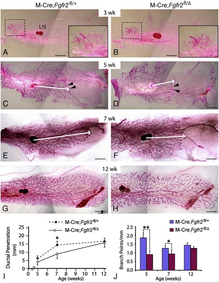
Fig. 2.
Consequences of MMTV-Cre-mediated inactivation of Fgfr2 in mammary epithelium. (A-H) The mammary branching tree at the postnatal stages indicated, as revealed by Carmine Red staining of glands in wholemount. (A, C, E, G) glands from control (M-Cre;Fgfr2fl/+) mice; (B, D, F, H) glands from mutant (M-Cre;Fgfr2fl/Δ) mice. Insets in panels A and B show high-magnification views of the rudimentary ductal tree (area in dashed box). (C-F) Arrowheads indicate TEBs at the tips of invading mammary epithelium. Arrows indicate the extent of ductal penetration in the fat pad. (I, J) Quantitative comparisons of ductal penetration and branch point formation between control and mutant glands. At 5 weeks, ductal penetration measurements were 6.0±1.4 (control, n=5) and 4.5±1.7 (mutant, n=6); at 7 weeks, the measurements were 14.6±2.2 (control, n=4) and 9.1±3.2 (mutant, n=4); at 12 weeks, they were 16.8±2.2 (control, n=8) and 16.0±2.9 (mutant, n=12). Measurements of branching points were 1.8±0.3 (control) and 0.9±0.2 (mutant) at 5 weeks, 1.3±0.1 (control) and 0.9±0.2 (mutant) at 7 weeks, and 1.4±0.1 (control) and 1.3±0.1 (mutant) at 12 weeks. Values shown are the mean±SD for each data point: **P<0.0005; *P<0.05, unpaired, two-tailed Student's t test. Scale bars: 2.5 mm. Abbreviation: LN, lymph node.

