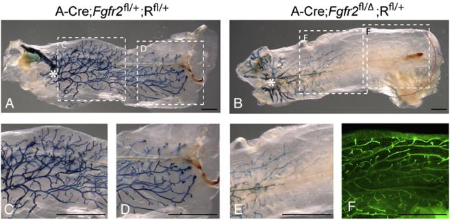
Fig. 4.
Analysis of Fgfr2 heterozygote/null genetic mosaics produced by Adenovirus Cre-mediated recombination. (A-E) Assays for β-GAL activity in mammary glands in which the ductal tree developed for 8 weeks from donor mammary epithelial cells harvested from (A) control Fgfr2+/+;Rfl/fl and (B) mutant Fgfr2fl/fl;Rfl/fl female mice and infected with adenovirus-Cre (see Materials and methods). Asterisks indicate the transplantation sites in the recipient fat pads from which donor cells grew out. Dashed boxes in panels A and B demarcate the regions shown at higher magnification in the corresponding panels (C-E). (F) The glands were stained with Yo-Yo1 to illuminate the branching tree; This staining, shown only for the region demarcated by the dashed box (F) in panel B, demonstrates that there are many epithelial branches in a region of the gland that contains virtually no β-GAL-positive/Fgfr2 null cells. 10 transplanted glands were examined for each genotype. Scale bars: 2.5 mm.

