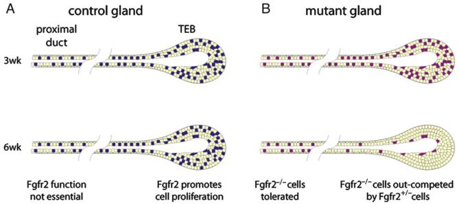
Fig. 6.
Model of FGFR2 function during postnatal branching morphogenesis in the mammary gland. (A) Schematic representations of primary branches in control genetic mosaic mammary glands at 3 and 6 weeks. Fgfr2 heterozygous cells are colored yellow and wild-type cells are colored blue. Fgfr2 is expressed in mammary epithelium, including TEBs and ducts, during postnatal development. Fgfr2 promotes cell proliferation of in TEB cells to ensure normal branching morphogenesis but is not required in the proximal ducts. (B) Mutant genetic mosaic mammary glands. Fgfr2 heterozygous cells are colored yellow and Fgfr2 null cells are colored purple. Fgfr2 null cells survive and persist in proximal ducts, but when TEBs undergo rapid cell proliferation and active epithelial invasion following the onset of puberty, Fgfr2 null cells are rapidly depleted. Once diluted out of the TEB, Fgfr2 null cells no longer contribute to the distal ductal network.

