Figure 1.
Detection of Nifedipine-Sensitive Tyrosine Phosphorylation Implicates Pyk2 in the GnRHa Signaling Network in αT3-1 Cells
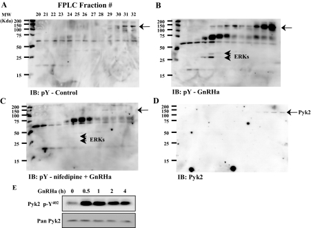
αT3-1 whole-cell lysates were fractionated by FPLC using an ion-exchange Mono Q column. Before fractionation, cells were treated with control solution (A), GnRHa buserelin (10 nm, 15 min; B), or nifedipine (2 μm) followed by GnRHa (nifedipine + GnRHa; C). Mono Q fractions 20–32 were resolved by SDS-PAGE and subjected to phosphotyrosine immunoblot analysis (IB: pY; A–C). The molecular markers are listed to the left of each panel. Short arrowheads identify tyrosine phosphorylation of ERKs in B and C. In D, blots were stripped and reprobed with antibody against Pyk2. Longer arrows identify tyrosine phosphorylation and colocalization with Pyk2 immunoreactivity. GnRHa treatment (100 nm) induced phosphorylation at Pyk2 Y402 as determined in whole-cell αT3-1 lysates over a 4-h time course (E) using a phosphospecific antibody and Western blot analysis. A Pyk2 antibody (Pan Pyk2) was used to demonstrate equivalent lane loading in E. MW, Molecular weight.

