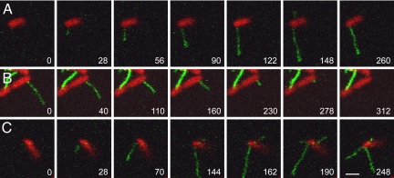
Fig. 1.
Extension and retraction of F-pili. (A) Pilus extension. The extension of a long pilus is seen in the first six frames; retraction is evident in the final frame. A second short pilus is visible only in the sixth frame. Note that during extension, the distal portion of a pilus is more brightly labeled than the cell-proximal portion, suggesting that the filament elongates from the base. (B) Pilus retraction. This 4-μm pilus retracted completely; during retraction fluorescence was uniform along the filament. (C) Independent regulation of F-pili on the same cell. This bacterium extended three pili, which grew and retracted asynchronously. For each bacterium, the complete time series may be seen as a movie: (A) Movie S1; (B) Movie S2; (C) Movie S4. Time is shown as seconds after the first frame. (Scale bar, 2 μm; all images are at the same magnification.)

