
Figure 1. Mice overexpressing ΔFosB exhibited biochemical markers of reduced dopamine signaling in the NAc.
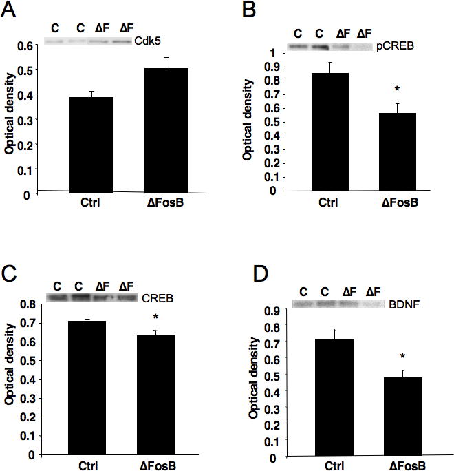
(A) ΔFosB mice displayed an indication of higher levels of Cdk5 than controls, similar to previous reports (Ctrl; P < 0.09). (B) pCREB levels were significantly reduced in ΔFosB mice compared to controls (P < 0.05). (C) Total CREB levels were also significantly reduced in ΔFosB mice (P = 0.05). (D) ΔFosB mice showed a significant reduction in BDNF levels in the NAc (P < 0.05). For all figures, representative bands are shown for control (C) and ΔFosB overexpressing (ΔF) mice. *Significantly different from control.