
Figure 5. High fat diet (HF) exposure and ΔFosB expression led to changes in expression of a number of key molecules in the VTA.
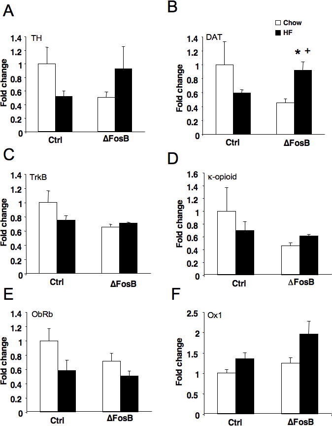
Expression levels were normalized to control (Ctrl) chow mice. (A) There was trend toward an interaction between diet exposure and ΔFosB expression on levels of tyrosine hydroxylase (TH; P < 0.06). HF diet decreased TH in control mice but increased expression in ΔFosB mice. (B) Expression of the dopamine transporter (DAT) was also differentially affected by both diet exposure and ΔFosB expression (P < 0.03). Similar to TH, DAT levels were decreased in control mice following HF diet and increased in ΔFosB mice. (C) Levels of the neurotrophin receptor TrkB showed a trend when corrected for multiple comparisons to be reduced in ΔFosB mice (P < 0.04), which appeared to be primarily due to differences in the basal state. (D) ΔFosB mice showed a trend for reduced expression of the κ-opioid receptor (κ-OR; P < 0.09). (E) Expression of the leptin receptor (ObRb) was significantly reduced following six weeks of HF diet (P < 0.03). (F) Expression of orexin receptor 1 (Ox1) was significantly increased overall following six weeks of HF diet (P < 0.02). *Significantly different from ΔFosB chow, +significantly different from control HF.