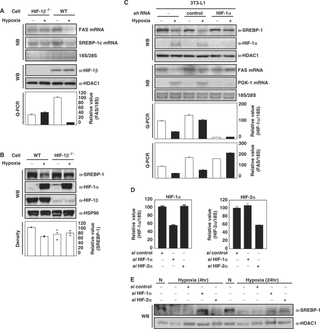
Figure 2.
Effect of HIF on hypoxic-repression of FAS and SREBP-1c. (A and B) Wild-type mouse Hepa1c1c7 cells and HIF-1β-defective Hepa1c1c7 cells were incubated in 1% O2 for 16 h. The levels of FAS and SREBP-1c mRNA were analyzed by NB and Q-PCR. WB analysis was performed using anti-SREBP-1 antibody. The level of SREBP-1 protein was estimated by measuring band intensities (LAS 3000, Fuji) and numbers represent averages and standard deviations of three independent experiments. WB with anti-HDAC1 antibody or anti-Hsp90 antibody were used as loading controls. (C) HIF-1α-knockdown 3T3-L1 cells and control 3T3-L1 cells were generated using the retroviral system as described in Materials and methods section. The cells were incubated in 1% O2 for 16 h. WB analyses, NB analyses and Q-PCR were performed. (D and E) Hepa1c1c7 cells were transfected with the indicated siRNAs as described. Before harvest, the transfected cells were exposed to hypoxia (1% O2, 4 h or 24 h). The mRNA levels were quantified by Q-PCR. Values represent means and standard deviations of three experiments.

