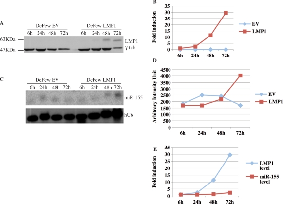
Figure 7.
Time-course of LMP1-mediated activation of miR-155. (A) Western blot analysis of protein extracts obtained at indicated times from DeFew cells infected with pBABEpuroLMP1 or empty vector. The blot was analyzed for the expression of LMP1 and normalized by γ-tub. (B) Graphic representation of the western blot results. (C) Northern blot analysis of RNAs obtained at indicated times from DeFew cells infected with retroviral vector expressing LMP1 or empty vector. The blot was analyzed for the expression of miR-155 and normalized by U6. (D) Graphic representation of the northern blot results. (E) Graphic representation of the correlation of miR-155 and LMP1 expression at indicated times.

