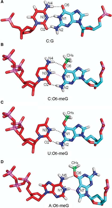
Figure 6.
Hydrogen-bonding interactions between (A) a G:C pair, O6-meG and (B) cytosine, (C) uridine and (D) adenine, respectively (see text). (A) G:C pair; G(O6):C(N4), G(N1):C(N3), and G(N2): C(O2), (B) anti,proximal O6-meG:C pair; O6-meG(O6):C(N4) and O6-meG(N2): C(O2), (C) anti,proximal O6-meG:U pair; O6-meG(N1): U(N3) and O6-meG(N2):U(O2) and (D) syn, distal O6-meG:A pair; O6-meG(O6): A(N6). Hydrogen bonds involving potential protonation sites, O6-meG(N1):C(N3+) and O6-meG(N7):A(N1+) are shown in pink. The methyl group carbon is colored in green. Color code is the same as in Figure 3. Details of the potential hydrogen-bonding partners are described in Supplementary Table S2.

