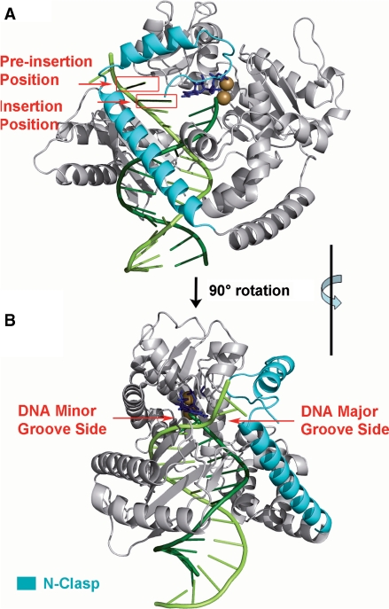
Figure 1.
(A) Definitions of preinsertion and insertion positions in the unmodified pol κ crystal structure (20) (PDB ID: 2OH2). (B) Denotation of DNA major and minor grooves shown through rotation of (A) by 90° along the vertical axis. Note that the N-clasp is on the major groove side. The enzyme is in gray with the N-clasp highlighted in cyan. The DNA template and primer strands are light and dark green, respectively. The incoming dNTP is blue. The Mg2+ ions are brown. Stereoviews are shown in Supplementary Figure S7.

