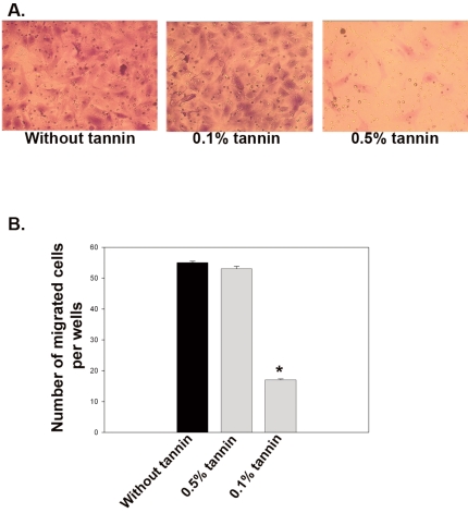
Figure 1.
Migration of rat carotid VSMCs after 10d of tannin treatment. Migration was stimulated by the addition of PDGF-BB (20 ng/mL) to the lower chamber. VSMCs were allowed to migrate for 14 h. Representative images of crystal violet-stained cells that migrated to the lower surface of the membrane are shown for tannin-treated (0.5% and 0.1%) vs untreated VSMCs (A). Cell counts on the lower face of the membrane reveal reduced motility of VSMCs treated with 0.1% tannin extract (B). The data are expressed as means +SD of each experiment performed in triplicate. *P<0.005 vs. untreated cells. n=4 wells per treatment group (4 fields/well).

