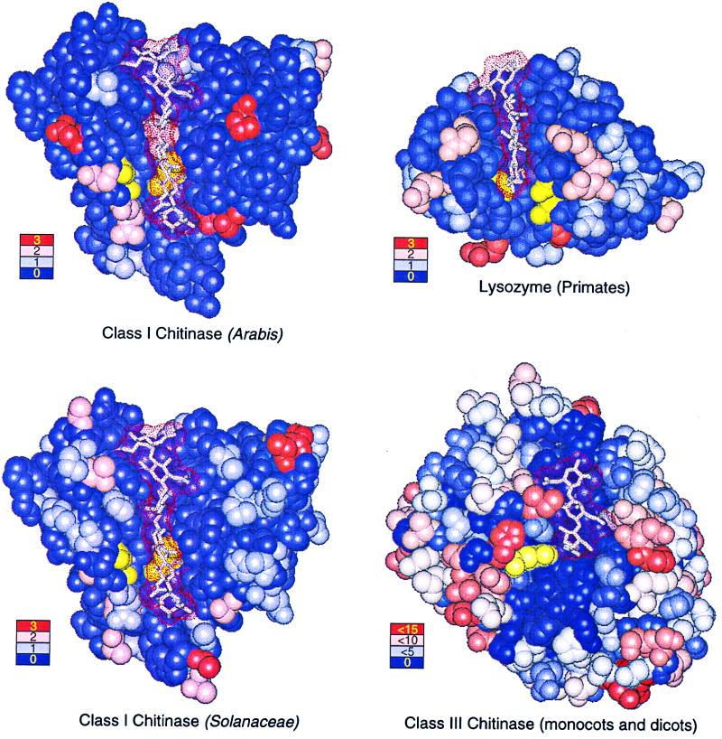
Figure 4.
Crystal structures with bound polysaccharide ligands (dot surfaces) showing the location and frequency of amino acid replacements for Arabis class I chitinase (Upper Left), Solanaceae class I chitinase (Lower Left), primate lysozyme (Upper Right), and plant class III chitinase (Lower Right). Ligands are hexa-N-acetyl-d-glucosamine (class I chitinases), tetra-acetyl-chitotetraose (lysozyme), and allosamadin inhibitor (class III chitinase). Catalytic residues are colored yellow. Color-coded legends show the number of replacements occurring at positions illustrated in the corresponding color.

