
Fig. 6.
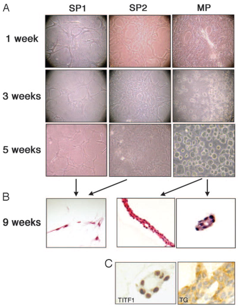
Morphological assessment of mouse thyroid SP and MP cells. SP1, SP2, and MP fractions were cultured using a three-dimensional collagen gel system, and cell morphology was recorded after 1, 3, and 5-wk culture (A). After 9 wk, cells were analyzed by hematoxylin and eosin staining for histology (B) and by immunohistochemistry (C) for TITF1 and TG. Only MP cells, but not SP1 or SP2 cells, developed epithelial arrangement and follicle-like structures that express TITF1 and TG. Magnification: ×100 (A), ×200 (B), and ×400 (C).