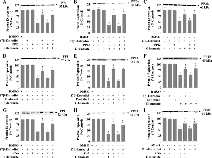
Figure 3.
PP1, PP2A, and calcineurin protein expression in response to glutamate and/or 17β-estradiol treatment in the presence of specific inhibitors of PP1, PP2A, or calcineurin in primary cortical neu. Primary cortical neurons were seeded in 100-mm dishes at a density of 500,000 cells/ml. Cells were treated simultaneously with 200 nm PPI2, 9 μm endothall, 500 nm cyclosporine A (CsA), 50 μm glutamate, and/or 100 nm 17β-estradiol. Cells were harvested after 24 h of treatment for Western blot analysis of PP1 (A, D, and G), PP2A (B, E, and H), and calcineurin (C, F, and I). Immunoblots were normalized to β-actin and graphs are represented as percent nontreated controls. Depicted are mean ± sem for six independent experiments. DMSO, Dimethyl sulfoxide. *, P < 0.05 vs. control; †, P < 0.05 vs. glutamate-treated group.

