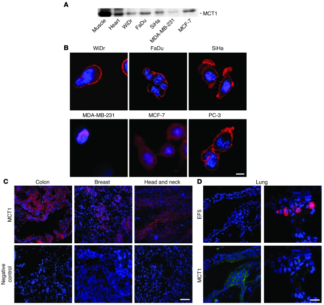
Figure 9. MCT1 is expressed in a variety of different human tumor cell lines and primary human tumor biopsies.
(A and B) MCT1 was detected by western blot (A) and confocal microscopy (B) in human tumor cell lines and control tissues. Note the plasma membrane expression of the lactate transporter in WiDr, FaDu, SiHa, and PC-3 cancer cells. (C) MCT1 (red) and nuclei (blue) were detected using immunofluorescence in biopsies of primary human colon, breast, and head and neck human cancers. (D) MCT1 and hypoxia were detected in cryoslices of a primary human lung cancer. The patient had received EF5 before tumor biopsy. Representative confocal microscopy pictures revealed that the staining of MCT1 (green) and of the hypoxia marker EF5 (red) did not overlap. Scale bars: 20 μm (B); 100 μm (C and D).

