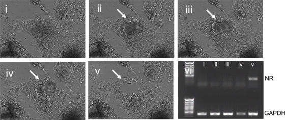
Figure 2. Automated aspiration of stem cell colony fractions from feeder cell monolayers.
Panel i.–v.: Automated documentation of a representative aspiration procedure using the CellCelector™/Soft Imaging System CellD. lane i.–v.: Determination of feeder cell contamination by RT-PCR expression analysis of nr mRNA.

