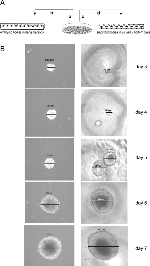
Figure 3. Embryoid body formation using standard conditions or feeder–freed stem cells.
A. Schematic representation of embryoid body formation: a. trypsination of stem/feeder cells, b. standard protocol for embryoid body formation in hanging drops, c. automated harvesting of feeder-freed stem cells colony fractions, d. transfer of feeder-freed stem cell to V-96 plate. B. Microscopic images of time dependent growth of (feeder-freed) f-f EBs and (standard) stEBs.

