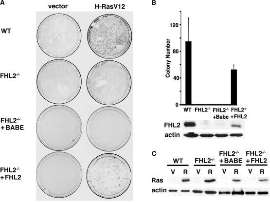
Figure 1. Absence of foci formation in immortalized FHL2−/− MEFs upon expression of H-RasV12.
(A) Focus formation assay of MEFs transiently transfected with H-RasV12 or empty vector. Plates were stained with Giemsa at 3 weeks after transfection. Data are representative of at least 3 independent experiments using 3 spontaneously immortalized clones. To restore FHL2 expression, primary FHL2−/− MEFs were infected with a retrovirus expressing FHL2 (pBabeFHL2puro) and cultured according to the 3T3 protocol. Two FHL2-restored FHL2−/− cell pools were obtained and used in the study. pBabe vector was used as a control (Babe). (B) Chart depicting at least three independent experiments. Mean colony numbers±standard deviation are shown. Bottom: Immunoblot analysis of FHL2 expression in FHL2-restored cell line. (C) Analysis of H-RasV12 expression in MEFs transfected with either empty (V) or H-RasV12 (R) expression vector. Samples were processed for immunoblotting 48 h after transfection. Actin was used as loading control.

