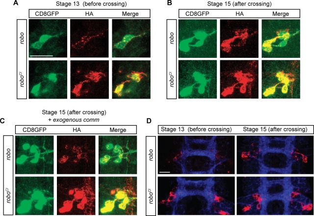
Figure 4. RoboSD is insensitive to Comm sorting in vivo.
(A–B) Confocal images of single cells expressing the fluorescent marker CD8GFP (green) and an HA-tagged version of either Robo or RoboSD (red). Panel (A) shows the soma of poxn-GAL4 expressing cells at stage 13 of embryonic development, namely while their growth cone is crossing the midline; panel (B) shows analogous cells at stage 15, after the crossing is complete. Notice that while there is no difference between Robo and RoboSD localization at stage 15 (B), a clear difference is observable at stage 13 (A). (C) Same experiment as in (B) but with ectopic expression of a uas-Comm transgene. (D) Lower magnification images of one segment of the CNS of UAS-HA-Robo (upper two) and UAS-HA-RoboSD (lower two) expressing embryos, before crossing (leftmost two) and after crossing (rightmost two). Images in panels A–C are acquired using 100× magnification; images in panel D are acquired using 63× magnification. White size-bar indicates 10 µm.

