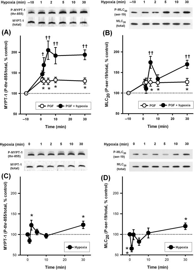
Figure 3.
Effects of hypoxia and PGF2α on phosphorylation of MYPT-1 (120–130 kDa) and MLC20 (18 kDa) in intra-pulmonary artery. (A and B) Phosphorylation at both sites was enhanced by 5 µM PGF2α (open circles, *P < 0.05 vs. control, n = 12–13 rats). In the continued presence of 5 µM PGF2α, hypoxia caused substantial further enhancement at both sites (filled circles, †P < 0.05, ††P < 0.01 vs. PGF2α alone, n = 12–17 rats). (C and D) In the absence of PGF2α, hypoxia caused a small sustained increase in phosphorylation (*P < 0.05, **P < 0.01 vs. control, n = 12–17 rats).

