
Figure 5. Failure of convergent extension in the definitive endoderm of chato mutants.
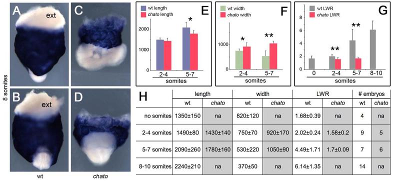
Wild type (A-B), chato (C-D) mutant 8 somite stage embryos hybridized with Ttr probes to highlight extraembryonic visceral endoderm (blue) and definitive endoderm (exterior layer of embryonic tissues in white). ext indicates white extraembryonic tissue. (A, C) lateral; (B, D) anterior views. (E) Plot of wild type (blue) and chato (red) definitive endoderm length. (F) Plot of wild type (green) and chato (red) definitive endoderm width. Data in μm. (G) Plot of definitive endoderm LWR in wild type (grey) and chato mutant (red) embryos. Error bars indicate standard deviation. * p<0.05; ** p<0.01 (H) Length and width average measurements ± standard deviation in μm. The number of embryos analyzed for each stage is indicated (# embryos). LWR, length to width ratio; na, not assayed.