
Fig. 4.
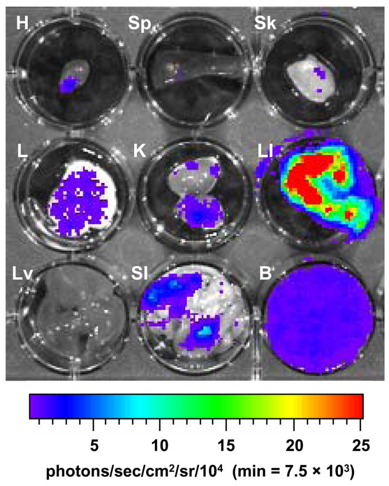
Detection of luminescent T. cruzi in the internal organs of infected A/J mice. Mice were infected with 104 trypomastigotes and injected with D-luciferin substrate (as described in Materials and methods) prior to sacrifice and organ dissection. Twenty-five days p.i. luminescence was analyzed in heart (H), spleen (Sp), skeletal muscle (Sk), lung (L), kidney (K), large intestine (LI), liver (Lv), small intestine (SI) and whole blood (B). For all images shown, the color scale ranges from blue (just above background with a minimum set to 7,500 photons/sec/cm2/sr) to red (maximum of 2.5 × 10⁵ photons/sec/cm2/sr). The minimum and maximum for this scale was adjusted to enhance signal detection while avoiding saturation and is consistent for all organs imaged. Abbreviation: min, minimum.