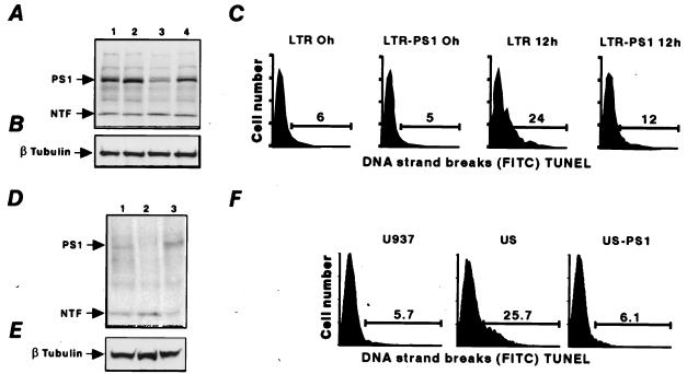
Figure 1.
PS1 is antiapoptotic. (A) Western blot analysis with anti-PS1 antibodies. (Lane 1) LTR6 cells at 37°C. (Lane 2) LTR6 cells transfected with PS1 (LTR-PS1) at 37°C. (Lane 3) LTR6 cells after 12 h at 32°C. (Lane 4) LTR-PS1 after 12 h at 32°C. PS1 indicates the full-length 50-kDa PS1 protein product, and NTF indicates the 30-kDa N-terminal fragment of PS1. (B) Western blot analysis with anti-β-tubulin antibodies. (C) Flow cytometry analysis of TUNEL assay in LTR6 cells and PS1 transfectants (LTR-PS1) at 37°C and after 12 h of incubation at 32°C. (D and E) Western blot analysis with anti-PS1 antibodies (D) or anti-β-tubulin antibodies (E) of protein extracts from U937 cells (lane 1), US cells (lane 2), and US cells transfected with PS1 (US-PS1) (lane 3). (F) Flow cytometry analysis of TUNEL assay in U937, US, and US-PS1 cells. Inhibition of apoptosis by PS1 is seen in both the LTR6-PS1 after 12 h and the US-PS1 transfectants.

